Steps for diagnosing lameness
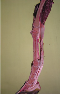
Cross-section forelimb.
Vets will develop their own protocols for assessing lameness, but the following steps give an indication of the process.
1. Review Medical History
The vet will ask you questions relating to past and present difficulties of your horse. They will also enquire about exercise or work requirements and any other pertinent information.
2. Veterinary Appraisal
While the horse is resting, the vet will study conformation, balance, and weight bearing and look for any evidence of injury or stress.
The vet will then observe your horse walking and trotting and, if appropriate, cantering and galloping. Assessment of the horse’s walk and trot will usually be done in hand, on a straight line over a firm, level surface.
The evaluation on different surfaces allows the lameness to be accentuated, which may give some suggestion as to the origin of the problem.
By observing the horse from the front, back and both side views, the vet will note any deviations in gait.
3. Hands-on Exam
The vet will palpate the horse, checking muscles, joints, bones and tendons for evidence of heat, swelling, tenderness or any other physical abnormalities.
Pulses in the blood vessels of the lower limb frequently yield useful information about infections or inflammation in this area.
4. Hoof Tests
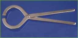
Hoof tester.
Hoof examination normally includes a careful visual inspection of the bearing surface of the foot when it is picked up.
By applying hoof testers to the sole, pressure is placed across specific areas of the hoof. Painful regions may be identified as the horse tries to retract its limb from the pressure exerted by the hoof tester.
5. Flexion Tests
Flexion tests help to assess the capsule surrounding joints together with the associated ligaments, tendons and bone ends. In an attempt to accentuate the lameness, the vet will hold the limb in a flexed position for a short period and then releases the leg. As the horse trots away the vet watches for signs of increased lameness.
However, as the response may be hard to interpret, a direct comparision should always be made with the opposite limb.
6. Lunging
Frequently the vet will want to watch the horse being lunged in a circle on soft or hard ground. This generally puts more pressure on the inside leg (front or back) and makes subtle lameness more obvious.
7. Alternative forms of Gait Analysis
Gait analysis or force plate analysis may be performed to observe hoof and limb placement.
As gait analysis is performed on a fast-moving treadmill, it is particularly useful when the lameness is only apparent at high speeds. Reflective markers are placed on the horse before it enters the treadmill and the procedure is videoed, thus allowing computerised analysis to monitor the motion of each joint.
Force plate analysis involves walking or trotting a horse over a sensor plate, which is sunken into the trot-up area. It measures the force applied through the placement of each hoof and allows the vet to determine if weight is being distributed abnormally.
8. Nerve and Joint Blocking
Once the affected limb has been identified and the lameness assessed, nerve and/or joint blocking should be performed to identify the area from which the pain arises. By using a specialist local anaesthetic to sequentially block the nerve impulses from the hoof upwards, it is possible to identify the location and source of pain.