Diagnostic imaging options
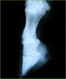
X-ray image of lower leg.
Once the area of pain has been identified, the following diagnostic imaging modalities can be used to identify abnormalities and obtain a diagnosis:
-
Radiography
A radiograph will show the internal structure of a horse. There are many conditions in which the actual extent of injury or disease can be accurately determined by X-rays, but the most important are the injuries and diseases of the bones.
-
Ultrasound
Ultrasonography is a means of diagnosing conditions of deep structures of the body by recording reflections of echoes produced by pulses of ultrasound waves.
-
Magnetic Resonance Imaging (MRI)
This technique is a hazard-free, non-invasive technique for generating images of internal sections of the body. The system works by utilising the differing absorption of radio waves by atoms in the body when exposed to a magnetic field. The amount of absorption is measured and the data used to generate a computer image.
-
Computerised topography
Topography is the systematic surveying, mapping, charting, and description of specific physical features pertaining to the injury.
-
Nuclear Scintigraphy
During nuclear scintigraphy (also known as a bone scan), the horse is injected with radioactive technetium, which binds to calcium in the bone. Where there is greater bone turnover, such as in diseased or damaged bone, then a greater uptake of radioactive technetium is seen. The resultant image may be used to identify the original location of the lameness.
Scintigraphy may be used:
- On occasions when the horse or situation will not allow other forms of assessment to be employed
- In the case of severe lameness
- When a lameness examination cannot be performed safely
- In the case of a lameness which cannot be blocked out by conventional nerve and joint blocks
-
Blood samples
Muscular problems can be identified by measuring the level of muscle enzymes in the blood.
Some diseases cause muscle pathology and therefore lameness. In these cases, it will not be possible to use nerve blocks to locate the source of pain. Instead, the use of blood analysis after exercise may show a marked elevation in muscle enzymes.